Products
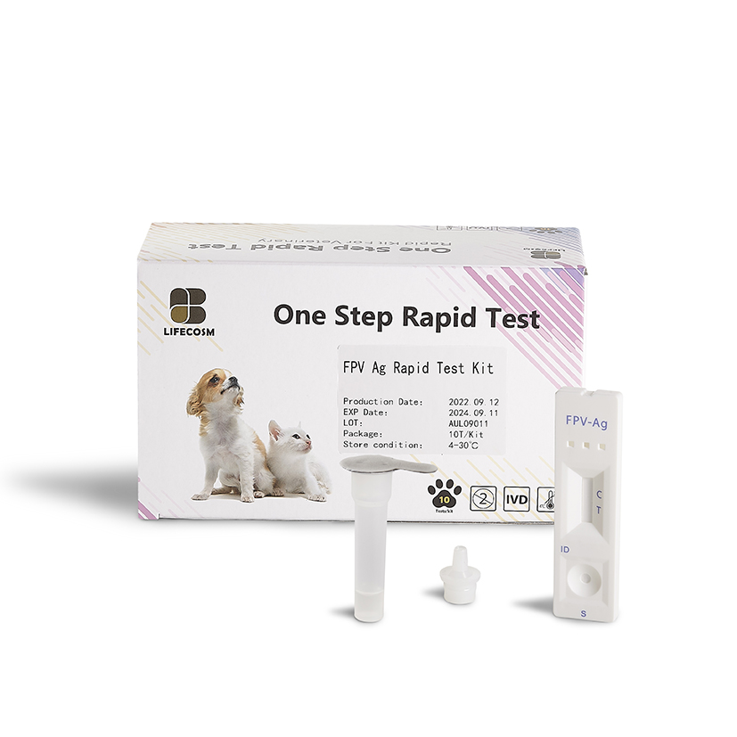
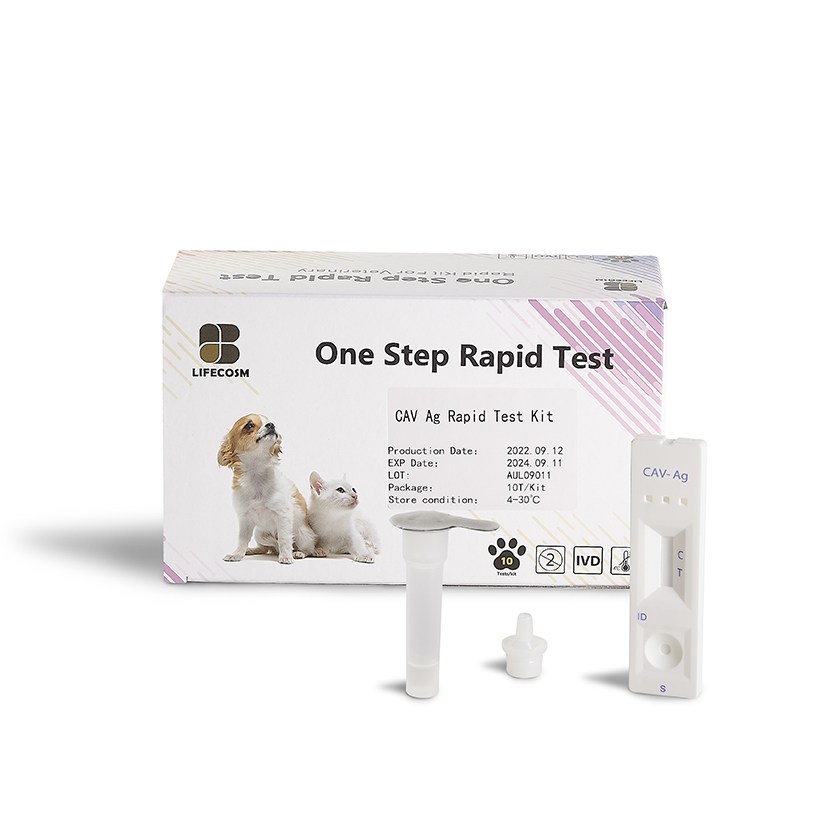
Lifecosm Feline Parvovirus Ag Test Kit
FPV Ag Test Kit
|
Feline Parvovirus Ag Test Kit |
|
| Catalog number | RC-CF14 |
| Summary | Detection of specific antigens of feline parvovirus within 10 minutes |
| Principle | One-step immunochromatographic assay |
| Detection Targets | Feline Parvovirus (FPV) antigens |
| Sample | Feline Feces |
| Reading time | 10 ~ 15 minutes |
| Sensitivity | 100.0 % vs. PCR |
| Specificity | 100.0 % vs. PCR |
| Quantity | 1 box (kit) = 10 devices (Individual packing) |
| Contents | Test kit, Buffer bottles, Disposable droppers, and Cotton swabs |
|
Caution |
Use within 10 minutes after opening Use appropriate amount of sample (0.1 ml of a dropper) Use after 15~30 minutes at RT if they are stored under cold circumstances Consider the test results as invalid after 10 minutes |
Information
Feline parvovirus is a virus that can cause severe disease in cats – particularly kittens. It can be fatal. As well as feline parvovirus (FPV), the disease is also known as feline infectious enteritis (FIE) and feline panleucopenia. This disease occurs worldwide, and nearly all cats are exposed by their first year because the virus is stable and ubiquitous.
Most cats contract FPV from a contaminated environment via infected feces rather than from infected cats. The virus may also sometimes spread through contact with bedding, food dishes, or even by handlers of infected cats.
Also, Without treatment, this disease is often fatal.
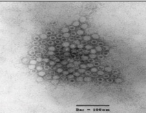
Symptoms
Ehrlichia canis infection in dogs is divided into 3 stages;
ACUTE PHASE: This is generally a very mild phase. The dog will be listless, off food, and may have enlarged lymph nodes. There may be fever as well but rarely does this phase kill a dog. Most clear the organism on their own but some will go on to the next phase.
SUBCLINICAL PHASE: In this phase, the dog appears normal. The organism has sequestered in the spleen and is essentially hiding out there.
CHRONIC PHASE: In this phase the dog gets sick again. Up to 60% of dogs infected with E. canis will have abnormal bleeding due to reduced platelets numbers. Deep inflammation in the eyes called “uveitis” may occur as a result of the long term immune stimulation. Neurologic effects may also be seen.
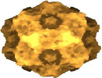
Diagnosis and treatment
In practice, FPV antigen detection in feces is usually carried out using commercially available latex agglutination or immunochromatographic tests. These tests have an acceptable sensitivity and specificity when compared to reference methods.
Diagnosis by electron microscopy has lost its importance due to more rapid and automated alternatives. Specialized laboratories offer PCR-based test on whole blood or feces. Whole blood is recommended in cats without diarrhoea or when no fecal samples are available.
Antibodies to FPV can also be detected by ELISA or indirect Immunofluorescence. However, the use of an antibody test is of limited value, because serological tests do not differentiate between infection- and vaccination-induced antibodies.
There is no cure for FPV but if the disease is detected in time, the symptoms can be treated and many cats recover with intensive care including good nursing, fluid therapy and assisted feeding. Treatment involves alleviating vomiting and diarrhea, to prevent subsequent dehydration, along with steps to prevent secondary bacterial infections, until the cat's natural immune system takes over.
Prevention
Vaccination is the main method of prevention. Primary vaccination courses usually start at nine weeks of age with a second injection at twelve weeks of age. Adult cats should receive annual boosters. The FPV vaccine is not recommended for kittens under eight weeks of age, since their natural immunity may interfere with the efficacy of the FPV vaccine.
Since the FPV virus is so hardy, and can persist in the environment for months or years, a thorough disinfection of the entire premises needs to be made after an outbreak of feline panleukopenia in a home shared by cats.

